CIRSE 2014: Phlebografic classification of anatomic variants in the right spermatic vein confluence: 23 years of work
Autori: Pieri Stefano, Agresti Paolo, Marzano Domenico
Financial disclosure
I have no financial disclosure or relationship with companies.
Purpose
Male varicocele is a clinical dysfunction, caused by a pathological venous reflux.
Knowledge of anatomic variants of the spermatic vein confluence is fundamental for the technical success of the percutaneous treatment.
While numerous articles have been written on the anatomy of the left spermatic vein, no exhaustive description exists for the controlateral vein.
Methods and materials
From a retrospective review of 5.885 patients treated for varicocele, with radiological intervention, between 1999 and 2011, we extrapolated phlebographic images and clinica data of patients with incontinence of the right spermatic vein only.
Indication for treatment was the presence of pain in the right inguinal region, in absence of a hystory of trauma, e/o seminal fluid alterations and evidence of venous reflux at eco-color-Doppler.
Phlebography have been performed only with a transbrachial approach, a multipurpose catheter and a hydrophilic guide wire and sodiotetradecilsolfato 3% for the sclerosis.
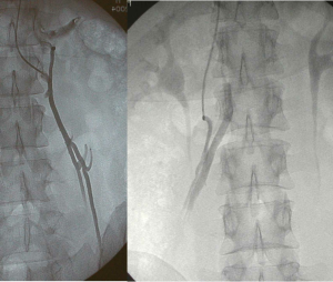
Contrast medium was manually injected, during Valsalva manouvre, in inferior vena cava and in the right renal vein [Fig. 1].
Fig.1 – Right vein phlebography during Valsalva manouvre; the internal right spermatic vein is visualized for the entire lenght, it appears uniformly dilatated and without valvular systems along its course. Very easy is the catheterization and the sclerosis
Results
We found 180/5.885 cases of incontinene of the right internal spermatic vein only (3,06%).
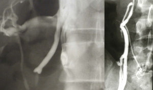
In the first group of patients (14 cases, 7,8%) the right spermatic vein drained exclusively into the renal vein, with one or more vessels [Fig 1 e 2];
Fig. 2 – In the first group of right spermatic vein confluence is possible to find a duplicated vein; during Valsalva manouvre is possible to visualize the entire lenght.
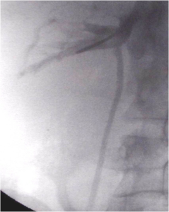
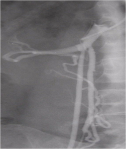
In the second group (42 patients, 23,4%) the vein drained into both the renal vein and the inferior vena cava, with one branch showing functional predominance.
Selective catheterization was easier to perform, on the first branch and confirmed dilatation of the vein as well as the absence of valvular systems.
 Fig. 3 – The spermatic phlebography from the confluence in the inferior vena cava shows another confluence, to the renal vein, bigger than the first, more easy to catheterize. Fig. 4 – In this case the right spermatic vein confluence is bigger into the inferior vena cava.
In most patients (124 cases, 65% ) the internal spermatic vein drained into the inferior cava vein : the confluence was double in 13 patients and single in the others.
Visualization of the incontinence was limited to the initial 5-10 cm of the vein in 26 cases; however, vein dilatation and absence of valvular systems were confirmed beyond the semicontinent valve.
 Fig. 5 – During inferior vena cava phlebography appears the confluence of the right spermatic vein [fig. 5] and a semicontinent valve. The catheterization was made with the hydrophilic wire first and the multipurpose after. [Fig. 6]
Conclusions
Interventional treatment is one of the therapeutics options for male varicocele, but the method is limited by the presence of anatomic variants or aberrantly supplying vessels which make catheterization and sclerosis of the internal spermatic vein difficult, if not impossible.
Interventional radiologists must have a through knowledge of anatomic variants of the right spermatic vein confluence, to be able to perform the procedure whitin a reasonable amount time and to reduce radiation exposure.

